Hibernomas, or pseudolipomas, are rare, benign soft tissue tumors derived from brown fat. The term hibernoma refers to the morphologic similarity of tumor cells to the brown adipose tissue (BAT) typically found in hibernating animals. BAT is present in newborns and is recognized as an important form of nonshivering thermogenesis in early life that significantly decreases in volume with age.
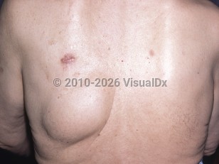
Hibernomas tend to present as painless, slow-growing masses that are firm, mobile, and nontender to palpation. They are well-defined, encapsulated, hypervascular lesions that typically range in diameter from 5-10 cm but may grow to larger than 20 cm. Hibernomas have no sex predilection and tend to occur in adults during middle age, although cases have been documented across ages. There are 4 distinct pathologic variants of hibernomas: typical, myxoid, spindle cell, and lipoma-like. Each variant can be found intermuscularly, intramuscularly, or subcutaneously. Variants are not readily distinguishable clinically or on imaging and require histology for distinction.
Hibernomas are not considered life-threatening and only manifest symptoms secondary to compression of adjacent structures. While often slow growing and asymptomatic, patients may present with rapid tumor growth, fever, and weight loss. There are no reported cases of malignant transformation, recurrence, or metastatic spread after complete excision. Accurate preoperative diagnosis is critical in reducing patient anxiety and guiding proper surgical and surveillance strategy.
There have been speculations about an association between hibernomas and multiple endocrine neoplasia type 1 (MEN1) as there have been a few case reports of hibernoma occurring in patients with MEN1.
Hibernoma
Alerts and Notices
Important News & Links
Synopsis

Codes
ICD10CM:
D17.9 – Benign lipomatous neoplasm, unspecified
SNOMEDCT:
404064001 – Hibernoma
D17.9 – Benign lipomatous neoplasm, unspecified
SNOMEDCT:
404064001 – Hibernoma
Look For
Subscription Required
Diagnostic Pearls
Subscription Required
Differential Diagnosis & Pitfalls

To perform a comparison, select diagnoses from the classic differential
Subscription Required
Best Tests
Subscription Required
Management Pearls
Subscription Required
Therapy
Subscription Required
References
Subscription Required
Last Reviewed:11/12/2020
Last Updated:11/12/2020
Last Updated:11/12/2020
Hibernoma