The genetic defect in PXE is a loss of function mutation in both alleles of the gene that encodes for the cellular transport protein ATP-binding cassette C6 (ABCC6). This transport protein appears to be an efflux pump for transporting ATP from hepatocytes, where it is then converted extracellularly to adenosine monophosphate (AMP) and inorganic pyrophosphate (PPi), which acts systemically as an inhibitor of mineralization of the elastic fibers. Prevalence is estimated to be from 1 in 25 000 to 1 in 100 000 with a slight female preponderance.
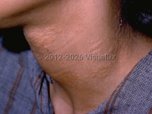
Clinical manifestations can be seen as early as the first or second decade of life. Cutaneous signs, the most frequent manifestation of the disease, appear as yellowish (xanthoma-like) papules on flexural areas. Commonly, the lateral neck is the first area of involvement, with other flexural areas (antecubital, popliteal, axillae, wrists, and groin) involved to variable degrees.
Angioid streaks (AS), which are breaks in Bruch's membrane due to calcification of the elastic lamina, are recognized as the most characteristic ocular finding in PXE. More commonly seen is a mottling peau d'orange retinal change representing subconfluent calcification of the membrane. Almost all patients will exhibit AS by the age of 30 years, and, while AS are themselves asymptomatic, choroidal neovascularity may develop in the fractures and lead to hemorrhage and subsequent scarring with gradual vision loss.
Cardiovascular manifestations are due to calcification of the elastic media and intima of vessels, primarily but not exclusively the medium-sized vessels of the extremities, resulting in premature atherosclerosis. Symptoms include intermittent claudication and angina pectoris. Patients often exhibit a decrease or loss of peripheral pulses, renovascular hypertension, and increased rates of mitral valve prolapse, myocardial infarction, and stroke. With involvement of the vessels of the gastric and intestinal mucosa, there is an increased incidence of gastrointestinal (GI) bleeding.
A PXE-like syndrome may also be precipitated by long-term D-penicillamine use for the treatment of cystinuria or Wilson disease.
Diagnostic criteria for PXE were proposed in 2014:
Major Criteria
Cutaneous:
- Characteristic papules and plaques on flexural areas
- Histopathology showing elastic fiber fragmentation and calcification in the reticular dermis
- Angioid streaks larger than one disc diameter in length
- Peau d'orange changes
- Molecular genetic testing for the presence of biallelic ABCC6 pathogenic variants
- PXE in a first-degree relative
- Genetic test with pathologic variant in one allele of ABCC6
- PXE changes in the histopathology of nonlesional skin
Probable diagnosis: An affected first-degree relative plus either both major cutaneous criteria OR one major ophthalmic criterion.