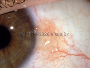
Conjunctival nevus - External and Internal Eye
Alerts and Notices
Important News & Links
Synopsis

Nevi of the conjunctiva are similar in pathology and behavior to those of the skin. They may be acquired or congenital, and they may be benign or malignant.
Pigmentation in congenital nevi may not be noted until adolescence.
Nevi may be intraepithelial, subepithelial, or compound. Spindle cell and blue nevi are less common. In puberty, intraepithelial nevi may mature and become more subepithelial, leading to a transition from flat to raised and a recognition of change from the patient.
Nevi are sometimes confused with benign acquired conjunctival melanosis or primary acquired melanosis, which is discussed in the section on conjunctival melanosis.
Codes
ICD10CM:
D31.00 – Benign neoplasm of unspecified conjunctiva
SNOMEDCT:
255006004 – Nevus of conjunctiva
D31.00 – Benign neoplasm of unspecified conjunctiva
SNOMEDCT:
255006004 – Nevus of conjunctiva
Look For
Subscription Required
Diagnostic Pearls
Subscription Required
Differential Diagnosis & Pitfalls

To perform a comparison, select diagnoses from the classic differential
Subscription Required
Best Tests
Subscription Required
Management Pearls
Subscription Required
Therapy
Subscription Required
References
Subscription Required
Last Updated:12/21/2008
Conjunctival nevus - External and Internal Eye

