Tinea pedis (athlete's foot) is a localized superficial fungal infection of the foot. Trichophyton rubrum, Trichophyton interdigitale, Trichophyton mentagrophytes, and Epidermophyton floccosum are the most common dermatophytes responsible for most cases of tinea pedis. Nondermatophyte molds and candidal yeasts can less frequently cause tinea pedis.
Tinea pedis is more common in men. The prevalence increases with age, with peak incidence between the ages of 15 and 45 years. Factors leading to this infection include high levels of humidity; occlusive footwear; and use of communal pools, showers, or baths (including locker rooms). Athletes are at increased risk (ie, "athlete's foot"). Infection can occur secondarily to close contact with infected individuals, animals (such as house pets), sharing clothing, or through fomites.
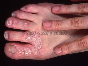
The clinical presentation of tinea pedis may vary. There are 3 main types of tinea pedis: interdigital (most common), hyperkeratotic / moccasin, and vesiculobullous tinea pedis.
In interdigital tinea pedis, the toe web spaces and soles of the feet are affected most frequently, with erythema, scale, and maceration of web spaces. The condition may spread to involve the nonplantar surfaces of the foot as well. Interdigital maceration, especially of the lateral fourth and fifth toe webs, is commonly seen.
Hyperkeratotic / moccasin tinea pedis presents with hyperkeratotic plaques with erythema on the lateral and medial foot, dorsum, and soles of the feet in a "moccasin" shoe pattern. There is often a collarette of scale along the border of the foot.
Vesiculobullous tinea pedis presents with pruritic and painful vesicles and bullae overlying the erythematous lesions.
Tinea incognito may occur when tinea pedis is misdiagnosed and treated with topical steroids, which reduces the scale and erythema of the lesions.
The infection may be pruritic or asymptomatic. It is often asymmetric, with only one foot being affected or more widespread on one foot than the other. In some cases, it can progress and cause concomitant onychomycosis (tinea unguium).
Secondary bacterial infections may occur, especially in diabetic patients, and are called dermatophytosis complex. Interdigital cracking and maceration may act as a portal of entry for pathogens and may predispose to lymphangitis or cellulitis. A dermatophytid reaction (also called an "id reaction") is a hypersensitivity process that can occur secondary to tinea pedis. The condition manifests on the lateral aspects of the fingers and may mimic dyshidrotic dermatitis, and may also be seen on the trunk and other parts of the body. This hypersensitivity process will resolve with adequate treatment of the dermatophyte infection.
Immunocompromised patient considerations: In patients with HIV infection and other immunodeficient states, interdigital tinea pedis has been noted to spread to involve the dorsal foot in an extensive manner.
Tinea pedis in Adult
See also in: Cellulitis DDxAlerts and Notices
Important News & Links
Synopsis

Codes
ICD10CM:
B35.3 – Tinea pedis
SNOMEDCT:
6020002 – Tinea pedis
B35.3 – Tinea pedis
SNOMEDCT:
6020002 – Tinea pedis
Look For
Subscription Required
Diagnostic Pearls
Subscription Required
Differential Diagnosis & Pitfalls

To perform a comparison, select diagnoses from the classic differential
Subscription Required
Best Tests
Subscription Required
Management Pearls
Subscription Required
Therapy
Subscription Required
References
Subscription Required
Last Reviewed:03/24/2025
Last Updated:03/27/2025
Last Updated:03/27/2025
 Patient Information for Tinea pedis in Adult
Premium Feature
VisualDx Patient Handouts
Available in the Elite package
- Improve treatment compliance
- Reduce after-hours questions
- Increase patient engagement and satisfaction
- Written in clear, easy-to-understand language. No confusing jargon.
- Available in English and Spanish
- Print out or email directly to your patient
Upgrade Today

Tinea pedis in Adult
See also in: Cellulitis DDx